
What is Medical Image AI?
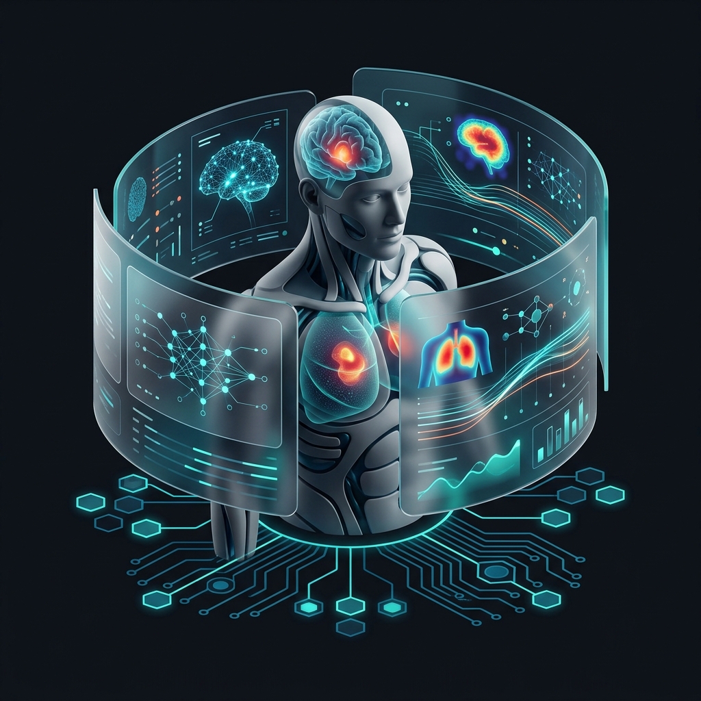
Medical image AI is a clinical technology that uses deep learning and computer vision to interpret diagnostic scans like CTs, MRIs, and X-rays. It identifies sub-visual patterns in high-dimensional pixel data, acting as an objective diagnostic partner for radiologists and specialists to improve accuracy and speed.
Medical Image AI is no longer a futuristic concept. In 2026, it is the backbone of modern clinical environments. From spotting micro-fractures that the human eye might miss to predicting cardiac events weeks in advance, artificial intelligence is rewriting the rules of patient care.
This guide explores the technical architecture, clinical applications, and implementation strategies for ai-driven diagnostics in the current healthcare era. Every hour of every day, these systems provide clinicians with a "second set of eyes" that remains objective, tireless, and hyper-precise.
- The evolution from rule-based systems to Agentic Medical AI
- Deep-dive into CNN foundation, Transformers, and GAN architectures
- Clinical applications across Radiology, Oncology, and Emergency Medicine
- A 4-phase roadmap for HIPAA-compliant AI implementation
- The role of Federated Learning and Multimodal AI in precision medicine
The Evolution of AI in Medical Imaging: From Rules to Reasoning
Automated medical image analysis is a method for extracting clinical insights from visual data. To appreciate where we are in 2026, we must look at how we got here. The first generation of medical imaging software relied on "rule-based" systems. These were simple algorithms that looked for specific thresholds. For example, a system might be programmed to flag a pixel if its Hounsfield Unit (HU) exceeded a certain level.
These systems were rigid. They failed to account for the massive variability in human anatomy. The second generation introduced "Machine Learning." Here, researchers manually selected features like shape or texture and fed them into a classifier. However, the true breakthrough came with the third generation: Deep Learning.
Instead of humans defining what to look for, the computer identifies its own features through a process of "auto-discovery." This changed everything. Today, in 2026, we are entering the fourth generation: Agentic Medical AI. These are not just static classifiers. They are autonomous agents. They can query a patient's history, compare current scans with historical data from three years ago, and suggest a complete diagnostic path.
"UseShift is at the forefront of this shift, helping clinics move from simple detection tools to fully integrated AI diagnostic partners. This is the step most teams skip — and it shows."

How AI in Medical Imaging Works (Technical Deep-Dive)
Automated medical image analysis is a method for processing high-resolution clinical scans using neural networks. To understand how these systems function, we must look past the user interface. At its core, the technology relies on neural network architectures designed to recognize patterns within high-dimensional data.
The Convolutional Neural Network (CNN) Foundation
The primary workhorse for deep learning for medical images is the Convolutional Neural Network (CNN). Unlike traditional algorithms that require manual feature engineering, where a human tells the computer what a "tumor" looks like, CNNs learn these features automatically.
- Convolutional Layers: These layers act as filters. They scan the image for edges, textures, and shapes. In medical imaging, the first few layers might detect the edge of a bone. Deeper layers detect the complex texture of a cancerous lesion.
- Pooling Layers: These reduce the spatial dimensions of the data. This makes the computation more efficient while retaining critical information. Efficiency matters when dealing with 8K resolution medical images.
- Fully Connected Layers: These layers perform the final classification. They determine if a specific pattern represents a benign cyst or a malignant growth.
Key Takeaways
- CNN-based models have achieved an Area Under the Curve (AUC) of 0.98 in identifying early-stage diabetic retinopathy.
- Transformers are replacing CNNs for full-body scans to understand long-range spatial relationships.
- GANs generate synthetic data for rare diseases and perform super-resolution upsampling.
Beyond CNNs: Transformers and GANs
While CNNs are excellent for spatial recognition, the industry is shifting toward Vision Transformers (ViTs). These models use "attention mechanisms" to understand the relationship between distant parts of an image. For example, in a full-body CT scan, a Transformer can correlate findings in the lungs with anomalies in the lymph nodes more effectively than a standard CNN.
Generative Adversarial Networks (GANs) are also playing a major role. They are used for data augmentation. They create synthetic, high-fidelity medical images to train models when real-world data is scarce or restricted by privacy laws. This is particularly useful for rare diseases where only a few hundred confirmed cases exist globally.
GANs are also used for Super-Resolution, taking a low-dose, noisy CT scan and "upsampling" it to a high-quality image that looks like it was taken with a much higher radiation dose. This improves clarity. Without increasing radiation exposure.
Dataset Quality and the Crisis of Reproducibility
A major challenge in artificial intelligence in radiology is "overfitting", where a model performs perfectly on the training data but fails in a real-world hospital. This often happens because the training data came from a single type of scanner or a specific patient demographic.
To combat this, developers are now using Cross-Institutional Validation. A model is trained at Hospital A but tested at Hospitals B, C, and D to verify generalizability. This process builds reliable validation frameworks, helping to confirm that ai-driven diagnostics remain accurate regardless of the hardware used.
The Role of DICOM and Data Annotation
For any automated system to work, it needs high-quality data. The industry standard is DICOM (Digital Imaging and Communications in Medicine). This format contains not just the image pixels, but also critical metadata like equipment settings and patient demographics.
Before a model is deployed, radiologists must "annotate" thousands of these files. They draw bounding boxes around anomalies or segment specific organs pixel-by-pixel. This "ground truth" data is what allows the AI to learn. Modern data pipelines now automate the preprocessing of DICOM files, which ensures that models are trained on clean, standardized data.

Key Applications: From Radiology to Oncology
AI-driven diagnostics is a method for automating the detection of pathologies in clinical imaging. The impact of these tools is visible across every major specialty. It is moving from a general-purpose tool to a highly specialized diagnostic partner.
Radiology
Automated triage of intracranial hemorrhage or tension pneumothorax. Flags life-threatening conditions to the top of the worklist.
Oncology
Radiomics for predicting treatment response and volumetric tumor segmentation accurate to the cubic millimeter.
Emergency
"Pan-Scan" CT analysis for trauma response using YOLO architectures for sub-60-second detection of organ lacerations.
Cardiology and Functional Imaging
Cardiologists use these tools to automate the calculation of the Ejection Fraction (EF) from echocardiograms. Traditionally, this was a manual, time-consuming process prone to inter-observer variability. AI provides a consistent, reproducible measurement in seconds. This allows for better management of congestive heart failure.
Dentistry and Orthodontics
The dental industry has seen a massive surge in AI adoption. Automated medical image analysis is used to detect caries (cavities), periodontal bone loss, and periapical lesions from 2D bitewings and 3D CBCT scans. This ensures that no treatment opportunity is missed.
Ophthalmology and Retinal Analysis
AI-driven retinal screening is now standard for managing diabetic patients. Portable cameras equipped with edge-AI can detect signs of retinopathy in primary care settings. This removes the need for every patient to visit a specialist for a routine check-up.
Comparison: Traditional vs. AI-Driven Medical Imaging
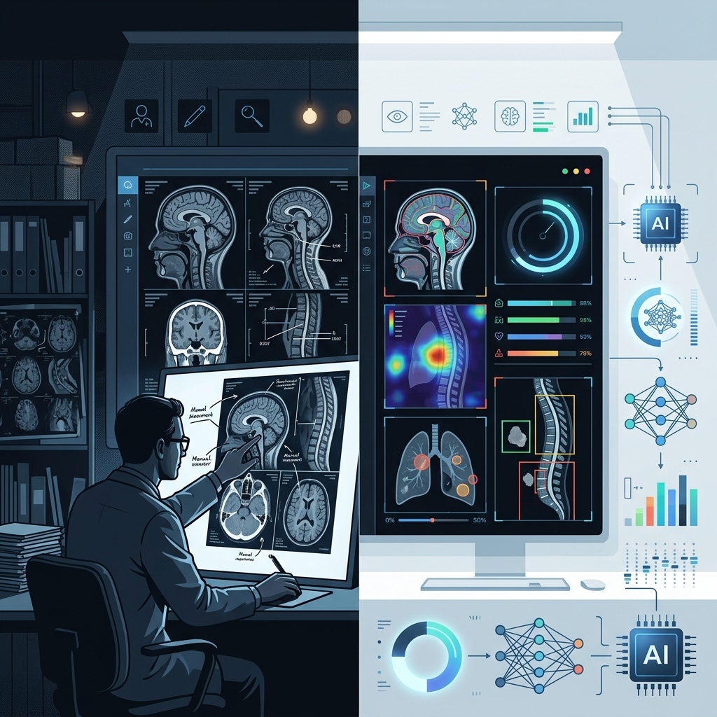
The shift toward ai-driven diagnostics represents a fundamental change in clinical efficiency. The following table highlights the key differences between the traditional manual workflow and the modern AI-integrated approach.
| Feature | Traditional Manual Analysis | AI-Driven Diagnostics (2026) |
|---|---|---|
| Analysis Speed | 10–30 minutes per complex scan | < 30 seconds for initial inference |
| Consistency | Subject to fatigue and bias | 100% consistent 24/7 |
| Early Detection | Relies on visible structural changes | Detects sub-visual textural patterns |
| Workflow | First-in, first-out queue | Intelligent triage (urgent cases first) |
| Data Usage | Limited to visual interpretation | Integrates with EHR and genomic data |
| Scalability | Limited by human headcount | Near-infinite horizontal scaling |
| Reporting | Manual transcription/dictation | Automated, structured reporting |
As the table demonstrates, these tools do not just do the same task faster; they change the nature of the task itself. By handling the rote work of detection and measurement, AI allows clinicians to focus on high-level decision-making and patient interaction.
Implementing AI Tools in Clinical Workflows
Successfully deploying automated systems in a clinical setting requires more than just a powerful model. It requires a seamless integration into the existing IT infrastructure.
The Implementation Roadmap: Phase 1 to Phase 4
Phase 1: Infrastructure Readiness Audit
Before installing software, you must assess your network bandwidth and server capacity. AI models, especially those processing 3D volumes like MRIs, require significant GPU power. You must decide between on-premise hardware or HIPAA-compliant cloud solutions.
Phase 2: PACS and RIS Integration
The Picture Archiving and Communication System (PACS) is the heart of any imaging department. For AI to be useful, it must live inside the PACS viewer. Integration ensures that AI findings appear as a native "AI Layer" in your existing workflow.
Phase 3: Pilot Program and Validation
Never roll out AI to the entire hospital at once. Start with a single department, such as the Chest X-ray queue in the ER. Compare the AI's findings with the senior radiologist's reports for 90 days. This "shadow mode" testing allows you to calibrate the AI's sensitivity and specificity.
Phase 4: Full Clinical Deployment
Once trust is established, the AI is moved to the frontline. At this stage, you should also automate the billing process. Over 45 specific CPT codes now exist for AI-driven procedures, ensuring that hospitals can receive direct reimbursement.
The "Black Box" Problem and Explainability
One of the biggest hurdles to adoption is the "Black Box" nature of deep learning. If an AI flags a lung nodule, the doctor needs to know why. Modern tools use Heatmaps (Grad-CAM) to highlight the specific pixels that triggered the positive result. This transparency builds trust.
Study Data: According to a 2024 study by the Mayo Clinic, clinicians who used "explainable" AI tools showed a 15% higher adherence to recommendations compared to those using "black box" systems.
Continuous Monitoring and Model Drift
AI models can degrade over time. This is known as "model drift." For instance, if a hospital upgrades its CT hardware, the new image characteristics might confuse an old AI model. Clinical teams must implement continuous monitoring to check that accuracy remains above established thresholds.
Multimodal AI: The Next Frontier
In 2026, the industry is moving away from "siloed" imaging. The most advanced systems now use Multimodal AI. This means the AI doesn't just look at the MRI; it also looks at the patient's latest blood work, their genetic markers, and their wearable device data.
For example, an AI might see a slight abnormality on a brain scan. On its own, it might be ignored. But when the AI sees that the patient also has a specific genetic predisposition for Alzheimer's and has shown a decrease in sleep quality over the last three months, it raises a high-priority alert. This is the essence of Precision Medicine.
The Role of Federated Learning in Healthcare AI
One of the biggest blockers to development is the difficulty of sharing sensitive patient data between institutions. Federated Learning (FL) solves this by bringing the AI model to the data, rather than the data to the model.
Standardized Federated Learning Workflow:
1. Distribute Global Model to Institutional Nodes
2. Local Training on Private DICOM Repositories
3. Aggregation of Weight Updates (Not Data)
4. Global Model Refinement & Redeployment
This process allows for the creation of massive, high-performance models without a single byte of patient data ever leaving the hospital's firewall. UseShift uses Federated Learning to help our clients participate in global research networks while maintaining absolute compliance with local privacy laws.
Regulatory Challenges and Ethical Compliance
The regulatory environment for deep learning for medical images is complex. In the United States, the FDA classifies these tools as Software as a Medical Device (SaMD).
FDA Approval Paths
Most AI tools enter the market via the 510(k) pathway, demonstrating that they are "substantially equivalent" to an existing legal device. However, for truly novel "autonomous" diagnostics, the De Novo pathway is required. By 2026, the FDA has streamlined these processes, focusing more on real-world performance data.
HIPAA and Data Privacy
Patient privacy is non-negotiable. Implementing AI requires strict adherence to HIPAA in the US and GDPR in Europe. This involves:
- Data Anonymization: Removing all Protected Health Information (PHI) from DICOM headers.
- Zero-Trust Architecture: Ensuring that only authorized clinicians can access AI results. Every access attempt is logged and audited.
Challenges of AI Adoption in 2026: The Clinician's Perspective
While the technology is ready, the "human factor" remains a challenge. Radiologists and specialists often express three primary concerns:
- Automation Bias: The tendency to stop checking the AI's work. If the AI says a scan is "clear," a tired doctor might skip the manual review.
- Alert Fatigue: If an AI flags too many "false positives," doctors will start to ignore the alerts entirely.
- Workflow Disruption: AI should not add more clicks to a doctor's day. Integration must be invisible and instantaneous.
The Ethics of AI-Initiated Diagnostics
As AI becomes more autonomous, ethical questions arise. If an AI misses a diagnosis, who is liable? The software developer? The hospital? The supervising doctor? In 2026, the legal framework is shifting toward Comparative Negligence, but the "Human-in-the-loop" model remains the gold standard for liability protection.
There is also the risk of bias in the AI's training data. If a model was only trained on images from one ethnic group, it might be less accurate for others. Ethical development requires "Diversity-First" data collection strategies.
Case Study: Mid-Size Regional Hospital Transformation
A 300-bed hospital in the Midwest integrated automated medical image analysis into their radiology department in early 2025. Before the integration, their "Turnaround Time" (TAT) for urgent ER scans was 45 minutes.
According to internal audit data (2025), after implementing an AI-driven triage system:
- TAT for Critical Cases: The turnaround time dropped to 12 minutes.
- Radiologist Burnout: Staff reported a 22% decrease in "cognitive fatigue" scores.
- Revenue: Annual revenue increased by $1.2M through improved throughput and specific AI-reimbursement codes.
This wasn't just about buying software. It was about shifting their operational model. They used automated frameworks to handle the data flow, allowing their doctors to do what they do best: save lives.
The Future of AI-Driven Healthcare
The trajectory of automated medical image analysis is clear: we are moving toward a future of Precision Medicine. In this future, your diagnostic scan is not just a picture of your body today. It is a roadmap for your health over the next decade.
The integration of AI into healthcare is not a "plug-and-play" operation. It requires a strategic shift in how data is managed, how clinicians work, and how patients are treated. Organizations that fail to adopt these clinical scanning tools will find themselves struggling with rising costs and declining patient outcomes.
UseShift is here to guide that transition. Whether you are a small clinic looking to automate your screening process or a large hospital system needing to integrate AI into your PACS, we provide the technical expertise to make it happen.
Ready to Scale Your Diagnostic Precision?
Deploy autonomous medical AI workflows that handle the heavy lifting, so your clinical team can focus on patient care.
Get a Free AI Strategy AuditKey Takeaways
- AI provides a 24/7 objective second set of eyes for clinical diagnostics.
- Technical shift from CNNs to Vision Transformers and Multimodal systems is defining 2026.
- Successful implementation requires deep integration with PACS/RIS and explainability frameworks.
Last updated: March 2026 | By Kristaps Jansons